
Orthodontics plays a vital role in ensuring that individuals have a healthy, functional, and aesthetically pleasing smile. Achieving the best results in orthodontic treatment requires a combination of skill, experience, and precise diagnostic tools. One of the most important tools in an orthodontist's diagnostic arsenal is X-ray imaging. Two types of X-rays commonly used in orthodontics are Cephalometric Radiographs (Ceph) and Panoramic Radiographs (Panorex). These X-rays provide critical insights that help orthodontists create effective treatment plans and monitor progress.
What Are Cephalometric and Panoramic X-rays?
Before diving into the importance of these X-rays in orthodontics, let’s define what they are:
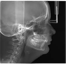
1. Cephalometric Radiographs (Ceph): These are side-view X-ray images of the head and face, showing the bones, teeth, and surrounding structures. The cephalometric X-ray is used primarily for analyzing the relationship between the teeth, jaws, and soft tissues. This type of X-ray is an essential tool for assessing skeletal relationships, determining the correct positioning of the teeth, and evaluating growth patterns over time.
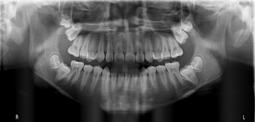
2. Panoramic Radiographs (Panorex): A panoramic X-ray captures a broad, two-dimensional view of the entire mouth area, including all teeth, the upper and lower jaws, sinuses, and other surrounding structures. Panoramic radiographs provide a comprehensive overview, making it easier to detect abnormalities in tooth eruption, jaw alignment, and the presence of impacted teeth or cysts.
Why Are These X-rays so Important in Orthodontics?
1. Comprehensive Diagnosis
One of the most critical aspects of orthodontic treatment is a thorough diagnosis. Both cephalometric and panoramic X-rays provide valuable information that cannot be obtained through visual examination alone. By using these images, orthodontists can assess the current condition of a patient's teeth, jaw, and bone structures, identifying any issues that may not be visible to the naked eye.
For instance, a panoramic X-ray allows orthodontists to see the full set of teeth, including those that are impacted or partially erupted. This is crucial for determining the appropriate treatment approach and anticipating potential complications.
2. Treatment Planning and Prediction
X-rays play an essential role in treatment planning. Cephalometric radiographs allow orthodontists to measure the size and position of the teeth and jaws. This information helps in formulating the most effective treatment plan, whether it involves braces, clear aligners, or other orthodontic appliances. The analysis of the cephalometric X-ray can also help predict how a patient's face and teeth will develop over time, allowing for more accurate and long-term planning.
Panoramic X-rays also aid in treatment planning, particularly when it comes to extractions. If a patient needs teeth removed to create space or correct alignment, a panoramic X-ray can help identify the best teeth to extract and ensure there are no underlying issues, such as cysts or tumors, that could complicate the procedure.
3. Monitoring Growth and Development
One of the significant benefits of using cephalometric X-rays is the ability to track and monitor a patient's growth and development. This is especially important for growing children and teenagers, where jaw and skeletal changes occur rapidly. Regular cephalometric X-rays allow orthodontists to evaluate how the facial bones and teeth are developing and adjust treatment plans accordingly.
For patients in their early teens, these X-rays are crucial for identifying potential problems like overbites, underbites, or crossbites and deciding the optimal time to start treatment for maximum effectiveness.
4. Assessing Bone Health
Both cephalometric and panoramic X-rays help assess the health of the bone structures. In orthodontics, proper bone structure is critical for the effective movement of teeth. Panoramic X-rays can help reveal bone loss or abnormalities, such as impacted wisdom teeth or jaw cysts, which could complicate orthodontic treatment if left untreated.
Cephalometric X-rays also help in evaluating the bone structure and relationships, which can guide orthodontists in determining if there are skeletal issues that need to be addressed, such as jaw discrepancies or malocclusions.
5. Planning for Surgical Intervention
In some cases, orthodontic patients may require surgery, especially if there are severe jaw discrepancies or skeletal issues that cannot be corrected through braces alone. Cephalometric X-rays provide detailed images of the skeletal system, which can be used to plan for surgical interventions. For example, when treating conditions like a severe overbite or underbite, orthodontists can use cephalometric analysis to determine the exact surgical approach needed and predict the results.